-
PDF
- Split View
-
Views
-
Cite
Cite
Emily Doole, A case of pancreatic pseudocyst as a rare cause of a cystic lesion within the psoas muscle, Journal of Surgical Case Reports, Volume 2021, Issue 11, November 2021, rjab499, https://doi.org/10.1093/jscr/rjab499
Close - Share Icon Share
Abstract
Acute pancreatitis is highly prevalent in Australia (Nesvaderani et al. Acute pancreatitis: update on management. Med J Aust 2015;202:420–3). Pancreatic pseudocysts, although typically occurring in the peripancreatic tissues, can in up to 25% be extra-pancreatic (Rasch et al. Management of pancreatic pseudocysts—a retrospective analysis. PLoS One 2017;12:e0184374). Extension of pseudocysts into the psoas muscle is highly unusual, with only 13 previously recorded cases (Gupta et al. Retroperitoneal cystic malignant fibromas mimicking a psoas abscess. Iran J Radiol 2015;12:e17507). This article presents the case of a 45-year-old man presenting with progressive left hip pain. He was known to have a history of chronic alcohol misuse. He presented with symptoms and signs typical of psoas pathology and was found to have a large pancreatic pseudocyst extending into his left psoas muscle. In this case, management was via both computed tomography guided percutaneous drainage and endoscopic ultrasound guided drainage. Because of the rarity of psoas pseudocysts and their propensity to mimic other pathology, diagnosis can be extremely challenging. Cystic lesions within the psoas muscle have several differentials, ranging from the more common psoas abscess to extremely rare neoplastic lesions.
INTRODUCTION
The worldwide incidence of acute pancreatitis is 4.9–73.4 per 100 000 people, with Australia sitting at the high end of this range [1]. Alcohol is the underlying aetiology for 70% of pseudocysts [2]. Seventy-five percent of pseudocysts are in the pancreas or peripancreatic tissues, most commonly the lesser sac [2]. The remaining 25% can be found in a wide range of extra-pancreatic locations, including mesentery, retroperitoneum, inguinal region, scrotum, liver, spleen, mediastinum, pleura and lung [4]. Extension into the psoas is extremely rare, with only 13 existing cases in the literature [3]. Other differentials of cystic lesions within the psoas muscle are broad and can be separated into neoplastic (both primary and metastatic), infective and other.
CASE PRESENTATION
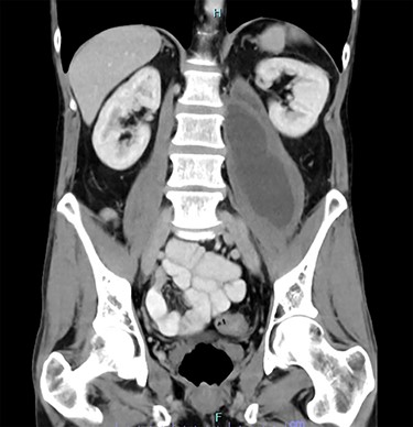
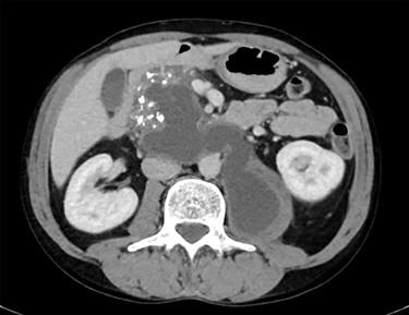
A 45-year-old man presented to his general practitioner complaining of 2 months of progressive left hip pain. Over the preceding 2 weeks he had been struggling to mobilize. His general practitioner (GP) investigated with a non-contrast computed tomography (CT) of the pelvis. This found a cystic lesion within the left psoas muscle with areas containing coarse calcification. The findings, overall, were suspicious of tumour, likely a teratoma. A CT chest, abdomen and pelvis with intravenous contrast was arranged. The pathology in the left psoas was found to represent a pancreatic pseudocyst extending from a heavily calcified pancreas, over the anterior aspects of the aorta and inferior vena cava (IVC) before reaching the left psoas muscle body (Figs 1 and 2). There were multiple other cystic regions noted within the pancreatic parenchyma and peripancreatic tissue. The patient was referred to the emergency department for surgical review.


CT showing calcified pancreas with pseudocyst extending from pancreas to left psoas muscle.
On examination of the patient in the emergency department the patient was lying in a supine position with his left hip in external rotation and left knee moderately flexed for comfort. He had an antalgic gait, with discomfort that was most evident weight bearing on his left leg. He also had pain elicited upon active movement of the left hip and in particular resisted hip flexion. He had no abdominal tenderness. Blood tests, including inflammatory markers and lipase, were within normal limits, with no evidence of acute pancreatitis.
The patient had a past medical history significant for a single episode of alcohol induced acute pancreatitis 10 years earlier. He had no diagnosed repeat episodes of pancreatitis in the intervening years. On further questioning he did mention occasional episodes of mild post-prandial epigastric pain. At the time of presentation, he had ongoing issues with alcohol misuse, consuming between 6 and 8 standard drinks per day.
On review of the CT scans, the dominant cyst was deemed not to be amenable to transgastric drainage. The psoas component of the cyst was however deemed appropriate for percutaneous drainage under CT guidance. Twenty millilitres of serous fluid was aspirated from the drain at the time of insertion, with minimal further output over the following 24 h. A repeat scan on Day 1 post drain tube insertion showed reduction in the size of left psoas collection. The drain tube was removed on Day 1.
Four weeks after his initial presentation the patient underwent endoscopic ultrasound and insertion of an AXIOS™ (Boston Scientific, Marlborough, Massachusetts, USA) stent. A 6-cm pseudocyst was identified in the pancreatic head, which was accessible from the second part of the duodenum and a 10-mm AXIOS™ stent was delivered into the pseudocyst. The stent remained in place for a total of 2 weeks and was then removed endoscopically.
At the time of writing, the patients’ symptoms had significantly improved following drainage of the dominant cysts. He does, however, have smaller pseudocysts remaining in the pancreas and peripancreatic tissues.
DISCUSSION
The differentials for cystic lesions within the psoas muscle are broad and can be categorized into neoplastic, infective and other. Primary malignant causes include teratoma, which was considered in this case, Schwannomas and primary soft tissue sarcomas with secondary cystic degeneration3. Several primary neoplasms have documented cases of psoas metastases including adenocarcinoma of the rectosigmoid, squamous cell carcinoma of the cervix and testicular tumours [4–6]. Infective causes include psoas abscesses and hydatid cysts [7]. Psoas haemorrhage or haematoma will also present as a cystic lesion [8]. Rarer documented causes include a case of an endometrioma embedded in the psoas muscle body, pseudo-abscesses containing calcium pyrophosphate crystals in a patient with pseudogout and a meningocele extended through the L1 neural foramina [9–11].
The diagnosis of pancreatic pseudocysts in extra-pancreatic locations can be a challenging one. In this case, the patient presented with classical signs and examination findings of psoas pathology with no known pre-existing conditions to lead towards aetiology. Extension into the psoas and surrounding tissues can also mimic other pathology, including inguinal hernias when presenting as a palpable groin lump, and even in one case complicated acute diverticulitis [12, 13].
The management of extra-pancreatic pseudocysts is difficult, as demonstrated in the highly variable management of the documented cases of psoas pseudocysts. Several were managed with imaging guided percutaneous drainage (ultrasound and CT), one with endoscopically guided drainage and even one with an open approach via laparotomy [12–15]. In the case above, due to the presence of numerous pseudocysts and loculations within the cysts several approaches were required to adequately control symptoms. Even with these approaches, there were several cysts remaining at the time of writing.
CONCLUSION
This case demonstrates both the complexities of diagnosing cystic lesions of the psoas muscle and extra-pancreatic pseudocysts. It also highlights the difficulties and variability in approach to management of pseudocysts extending into the psoas muscle.
AUTHOR’S CONTIBUTIONS
ED collected case information and was directly involved in patient care, collated patient information and formulated the review of literature, case review and discussion. The corresponding author attests that all listed authors meet authorship criteria and that no others meeting the criteria have been omitted.
COMPETING INTERESTS
The authors have no competing interests to declare.
FUNDING
This research did not receive any specific grant from funding agencies in the public, commercial or not-for-profit sector.
ETHICAL APPROVAL
Informed written consent was obtained from the patient included in this case report.
DATA SHARING
Data sharing is not applicable to this article as no datasets were generated or analysed during the current study.


