


Abstract
Klippel-Trénaunay-Weber syndrome (KTWS) is a rare condition characterized by a classic clinical triad. However, it can also have other features, such as cavocarus foot deformity, which is a rare presentation in a patient with KTWS. In this case report, we present our surgical technique of correcting such a complex deformity. Also, there are no other similar cases reported in the literature. An 18-year-old girl who is a known case of KTWS with a complaint of progressive unilateral left foot deformity. Her examination, revealed a rigid pes cavus with an equinus deformity in the left foot. Radiography of the left foot revealed marked cavus as well as hindfoot and forefoot varus. A successful surgical correction of the deformity was described in a stepwise fashion. The management of foot cavocarus deformity in KTWS patients is associated with high intra- and post-operative risk due to its complexity. Therefore, the management requires a multidisciplinary team approach.
INTRODUCTION
The International Society for the Study of Vascular Anomalies’ 2014 classification of vascular anomalies defines Klippel-Trénaunay-Weber syndrome (KTWS) as a complex rare congenital syndrome with a triad of capillary and venous malformations as well as limb overgrowth with or without lymphatic malformation [1]. The incidence of the syndrome is about 1:100 000 and has no association with gender, race or geographical area, and occurs sporadically [2]. The exact etiology and pathogenesis of KTWS are obscure. However, most of the cases were caused by mosaic-activating mutations in the PIK3CA gene [3–6]. Most cases of KTWS involve one extremity. However, 70 patients had involvement of bilateral extremities and were reported by Jacob et al. [7] Although the leg is the most commonly affected site, the site of involvement could differ from one patient to another. Almost two-thirds of patients with KTWS present with orthopedic manifestations [2]. The reported orthopedic conditions associated with KTWS are limb-length discrepancy, scoliosis, kyphoscoliosis, syndactyly, angular deformity and limb/joint pain [8].
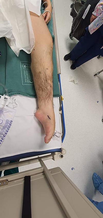
Side photo for the left lower limb showing severe cavus of the foot.
In this presentation, we report our surgical management of a patient with complex foot deformity in the form of progressive equino-cavovarus. Our main goal of this study is to describe our surgical technique in treating such deformity in a stepwise fashion.
CASE HISTORY
An 18-year-old girl with KTWS presented with progressive equino-cavovarus deformity of the left foot. The patient was in her usual state of health until 5 years ago, when she started to complain of left leg fatigue and pain when she walked for long distances. Two years later, she started to notice progressive deformity in her left foot, which increased her walking difficulty to the extent that she walked with a cane. There was no history of pain at rest, trauma, numbness or other neurological symptoms. She had an arteriovenous malformation (AVM) in the left lower limb on the anterolateral distal third of the thigh since birth. The AVM was stable, did not increase in size or cause any other problems.
Examination of the left lower limb revealed hemihypertrophy and superficial dilated vessels. There was a rigid pes cavus with equinus deformity in the left foot (Fig. 1). Neurological examination revealed the power of left foot dorsiflexion/plantarflexion was zero out of five and inversion/eversion was two out of five. Reflexes, tone and sensations in both limbs were normal. The vascular exam showed palpable dorsalis pedis and femoral pulses bilaterally. It was difficult to palpate left popliteal pulses. The posterior tibial pulse was absent bilaterally. The right foot examination was unremarkable otherwise.
Magnetic resonance imaging (MRI) brain and whole spine were ordered for her due to the neurological pattern of the disease. The MRI showed T4–T5 posterior fusion, there were no other neurological lesions. For evaluation of the varicose veins, vascular consultation was obtained, and Doppler ultrasound was ordered, which showed absent popliteal deep veins. Radiography of the left foot revealed marked cavus as well as hindfoot and forefoot varus. Also, it showed soft tissue swelling, reduction in bone density, and no evidence of fracture or dislocation or tarsal coalition (Fig. 2). The right foot radiograph was unremarkable apart from mild hallux valgus deformity (Fig. 3).
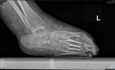
Lateral weight bearing radiograph showing severe pes cavus with osteopenia and soft tissue swelling.
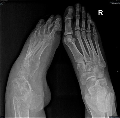
AP radiograph of both feet showing cavovarus deformity of left foot and right hallux valgus.

Lateral foot radiograph showing correction of the deformity after calcenus osteotomy.
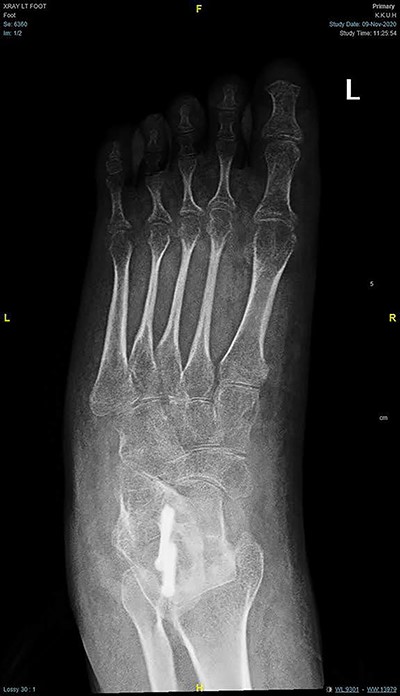
AP radiograph of the foot showing corrected for foot adduction.
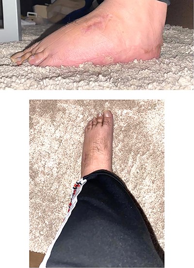
Side and front photo for the left lower limb postoperative (after 6 months).
After discussing the surgical intervention with the patient and her family, she opted for the surgical management due to the progressive nature of the deformity, which affected her ability to walk.
In the operation room, the patient was placed prone, then supine on a radiolucent table under general anesthesia. A tourniquet was placed below the fibular head without Esmark. Tourniquet compression was suggested by the vascular surgeon due to her deep vein malformations. We started our procedure with a standard posteromedial approach. Percutaneous Achilles tendon lengthening by triple hemisectiontendon and isolation of the posterior tibial tendon were done. Another medial ankle approach was done to incise the posterior tibial tendon from its insertion at the navicular. Extensive venous plexus was noticed in the midfoot, which was isolated and protected. Posterior tibial tendon transfer anteriorly through the interosseous membrane. Lateral sliding calcaneal osteotomy was done through lateral calcaneal approach and fixed with two 5.0 mm cannulated headless screws under image guide (Figs 4 and 5). In addition, two incisions were done at the dorsum of the midfoot and the anterior distal one-third of the left leg corresponding to the reflected posterior tibial tendon from posterior to anterior. The tendon was then passed under the extensor retinaculum and delivered in the dorsum of the midfoot. Sutures anchors were used to fix the tendon in the lateral cuneiform. Finally, planter release was done for all five toes through snip penetration with 11 blades. The overall correction was found acceptable intraoperatively. All incisions were then closed in layers. Then, she was placed on a controlled ankle motion boot (CAM boot) for 3 days to allow for swelling, followed by a U-shape and backslap splint. She was kept non-weight bearing for 6 weeks to allow for bone healing. After 6 weeks, the protocol was advancing the weight-bearing status from non-weight bearing to partial to full weight-bearing as tolerated with the CAM boot. Moreover, gait and range of motion exercises were started around the tenth week. The patient reported significant improvement in her ability to walk and overall satisfaction with the surgery at the fourth month postoperative (Fig. 6).
DISCUSSION
Our case report is an 18-year-old girl with KTWS who complained of progressive Equino-cavovarus of the left foot. In the literature, there are no other similar cases reported.
Most cases of KTWS involve one extremity. However, 70 patients had involvement of bilateral extremities and were reported by Jacob et al. [3] Although the leg is the most commonly affected site, the site of involvement could differ from one to another. With one or more of the triad features, patients may be diagnosed with KTWS [4]. Almost two-thirds of patients with KTWS present with orthopedic manifestations [2]. Therefore, an orthopedic surgeon is crucial, and it is recommended to involve him early to guide the optimal timing of both surgical and non-surgical intervention. In the literature, KTWS and its associated foot anomalies have been described infrequently [9–11].
In conclusion, managing equino-cavovarus in patients with KTWS is a complex problem and needs good operative planning. Our main goal of this study is to describe our surgical technique in treating such deformity in a stepwise fashion. Extensive hemorrhage and delayed bone healing are the main operative complications. These complications can be prevented by pre-operative optimization and good nutrition. While our patient recovered well and showed improved function with less trouble walking, the follow-up time for a final outcome is relatively short.
CONSENT
The patient has provided written consent for the publishing of the report.
ETHICAL APPROVAL
The ethical committee of King Khaled University Hospital has be requested to except this case report from obtaining an ethical approval.
AUTHORS’ CONTRIBUTIONS
The first and the second contributor prepared the first draft of the manuscript and treated the patient. All contributors conducted a literature search, contributed to preparation of the manuscript and prepared the final manuscript.
CONFLICT OF INTERESTS STATEMENT
The authors declare that there is no conflict of interests regarding the publication of this paper.
FUNDING
None.
