


Abstract
A 66-year-old man underwent a minimally invasive oesophagectomy for oesophageal adenocarcinoma. Surgery and recovery were routine; however, he represented 8 days later with a massive upper gastrointestinal bleed. He was stabilized, but over a 2-week period experienced several bleeds requiring transfusion and multiple endoscopies, all showing a prominent luminal vessel at the oesophago-gastric (OG) anastomosis. Haemostatic clipping was attempted resulting in pulsatile bleeding and transfer to the radiology suite where angiography showed extravasation of contrast at the OG anastomosis from the terminal portion of the gastro-epiploic arcade. Coil embolization was successful and did not result in ischaemia. It was our standard to construct the OG anastomosis with the end-to-end anastomosis circular stapler (DST™ Series EEA™), 4.8-mm staple height. However, we now use the 3.5-mm staple height for improved haemostasis and ensure that the area for anastomosis is cleared of omental tissue so as not to incorporate a visible vessel.
INTRODUCTION
One of the critical steps of an oesophagectomy is ensuring that there is adequate blood supply to the gastric conduit and the oesophagogastric (OG) anastomosis. Delayed bleeding at the anastomosis is rarely reported in the literature [1, 2]. Here, we present such case and the management difficulties this presents.
CASE REPORT
A 66-year-old man was referred to our service with the diagnosis of oesophageal adenocarcinoma. He had a complete preoperative workup and was deemed to have localized disease with clinical stage of T3N0M0. Past-medical history included a minor stroke, a deep vein thrombosis 20 years prior treated with lifelong anticoagulation, and type 2 diabetes. He underwent neoadjuvant chemo-radiation followed by a minimally invasive ivor lewis oesophagectomy. As is our standard, the OG anastomosis was created using an end-to-end anastomosis (EEA) circular stapler (DST™ Series EEA™ 25-mm Staple, 4.8-mm staple height; Medtronic, Minneapolis, MN) utilizing the OrVil™ device (Medtronic, Minneapolis, MN). His recovery was routine with a normal radiological swallow test on day 5 and discharge on day 12.
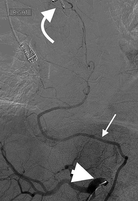
Angiography of the right gastro-epiploic artery. Arrowhead = celiac trunk; Arrow = right gastro-epiploic; Curved arrow = OG anastomosis.
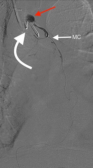
Angiography showing extravasation at the anastomosis. Curved arrow = OG anastomosis; Red arrow = contrast extravasation; Arrow MC = microcatheter.
Eight days later, he represented with an upper gastrointestinal bleed with massive haematemesis requiring immediate intubation for airway protection. Computer tomography angiography was unremarkable and an emergency gastroscopy revealed an extensive amount of blood and clot in the oesophagus and gastric conduit (limiting the view), but no active bleeding point. Conservative treatment in the Intensive Care Unit (ICU) with blood transfusion, proton pump inhibitors and life support were implemented. Another large bleed occurred 2 days later and a repeat gastroscopy revealed a non-bleeding visible vessel at the OG anastomosis and a haemostatic clip was applied. However, over the next 10 days, the patient remained blood transfusion dependent and had two further endoscopies. The first was unremarkable, but the second endoscopy re-demonstrated the luminal vessel at the OG anastomosis, with no evidence of the previously placed clip. This was felt to be the cause of his intermittent and problematic bleeding, so another haemostatic clip was attempted, resulting in pulsatile, moderate volume arterial bleeding. The procedure was abandoned and the patient transported immediately to the Interventional Radiology Suite. Right femoral access was obtained and angiography performed. The thoracic aorta was normal, so the celiac trunk was cannulated and angiography of the right gastro-epiploic artery performed (Fig. 1), revealing active extravasation of contrast at the OG anastomosis as shown in Fig. 2. A microcatheter was advanced along the length of the right gastro-epiploic artery to the OG anastomosis and coil embolization was performed (Fig. 3). Care had to be taken to ensure that the embolization was as distal as possible to limit the risk of ischaemic complications. The patient required lengthy ongoing care in the ICU for organ dysfunction but remained hemodynamically stable after embolization without evidence of conduit ischaemia. His hospital stay was obviously lengthy, but he was ultimately transferred to a rehabilitation unit.
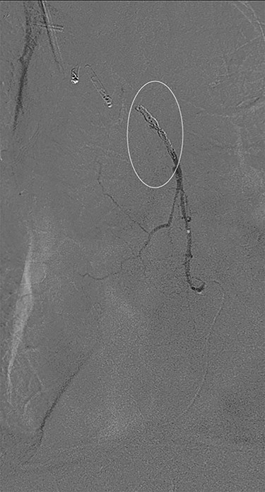
Successful coil embolization of the terminal portion of the gastro-epiploic arcade. Ellipse = coil.
DISCUSSION
Oesophagectomy is a major operation with significant perioperative morbidity. Respiratory and cardiac complications, along with gastric conduit ischemia and anastomotic leak, tend to dominate [3]. However, bleeding complications apart from those occurring intra-operatively are rarely reported [1, 2]. We present a unique case of an anastomotic bleed from a terminal branch of the gastro-epiploic vascular arcade. While the origin of this vascular arcade is a defined vessel (right gastric-epiploic artery) visible to the naked eye, the terminal portion is comprised of submucosal arterioles and capillaries and often devoid of a visible vessel [4]. However, as shown in Fig. 1, vascular supply to the terminal portion of the gastric conduit is present and can be abundant. The bleeding in this case likely occurred from incorporation in the anastomosis of either an invisible submucosal arteriole or the terminal portion of the right gastro-epiploic artery hidden within omental tissue. It is our standard practise to construct the OG anastomosis with the EEA circular stapler. It is common surgical knowledge that the oesophageal wall is thick [5] and has been our practise to use the 4.8-mm EEA staple height. There are several descriptions in the literature about this anastomotic technique and most recommend this staple height [6–8]. However, we believe that the higher staple height may have contributed to the bleeding issue in this case as there may have been insufficient compression of a submucosal arteriole. While it is impossible to be certain if a lower staple height would have prevented this situation, this case has resulted in our change to use the 3.5-mm EEA. In addition to this, we have become more meticulous in ensuring that the portion of the great curvature to be used for the anastomosis is cleared of excessive omental tissue to be sure no visible vessel is present that may get caught in the anastomosis.
References
Last R, McMinn R (ed). Thorax. In: Last’s Anatomy: Regional and Applied, 9th edn. Churchill Livingstone, Robert Stevenson House, 1-3 Baxter's Place, Leithwalk, Edinburgh, UK. 1994, pp 278.
