


Abstract
Complications following spinal surgery can range from simple wound infection to complete paralysis. Intraoperative checks have been introduced to account for all the instruments and materials used and help minimize surgeon-related complications. We report a case of a broken osteotome tip within the spinal canal following a routine posterior decompression of the lumbar spine.
INTRODUCTION
There have been great emphasis and wide improvements in the surgical management of the patient following the World Health Organisation surgical safety checklist. Prior to closure of the surgical wound, numerous steps and checks are implemented to ensure that the correct number of swabs and instruments are present with the scrub nurse to prevent any catastrophes, which to this day still occasionally occur. When the instrument count is complete, the surgeon can complete the closure. This report highlights the importance of scrutiny of each individual instrument especially when used in spinal surgery as complications can often lead to devastating complications.
CASE REPORT
A 68-year-old male presented with back pain and left-sided sciatica associated with ipsilateral foot drop. An MRI scan revealed severe lumbar degeneration at the level of L3/4 and L4/5 with significant central canal stenosis. He underwent an elective posterior lumbar spinal decompression with no instrumentation.
Day 1 postoperatively, the patient complained of reduced sensation in the L4 and L5 dermatomes on the right side. On examination, he had a right foot drop. Day 2 postoperatively, scrotal sensation and anal tone were both found to be reduced.
Postoperative Anterior-Posterior and lateral (Figs 1 and 2) radiographs of the lumbo-sacral spine revealed a radio-opaque foreign body on the right side of the spinal canal. The patient returned to theatre 3 days following the original operation for wound exploration. The foreign body was found to be lying in the spinal canal on the right side with an associated small dural tear. The object was identified as a broken metal tip and traced to an osteotome used in the primary procedure.
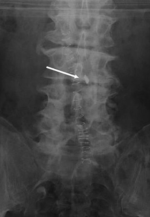
Postoperative plain Anterior-Posterior film.
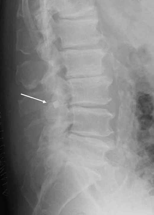
Postoperative plain lateral film.
DISCUSSION
The most plausible explanation for this complication is that during the release of the hypertrophied facet joint, the osteotome tip broke. What is particularly concerning is the fact that although the approach was on the left side, the broken tip was able to traverse the midline and settle adjacent to the spinal canal on the right, tearing the dura in the process.
With an uneventful procedure at the time and routine intraoperative counts being correct, there was no obvious reason for scrutinizing the standard instrument set used.
Romero et al. [1] assessed the use of routine radiographs during 670 visits in 202 patients who underwent posterior instrumented lumbar spine fusions. Only one patient visit in 594 radiographs had radiographic findings that prompted additional action. They concluded that routine radiographs had limited use in the early postoperative period after instrumented lumbar spine fusion [2]. Likewise, Grimm et al. did not find any use for routine postoperative radiographs after cervical spine fusions [3].
Postoperatively, no radiographic imaging of the spine was undertaken until the patient presented with abnormal signs and symptoms. Within our centre, as a result, routine postoperative spinal radiographs are only done following implantation of devices for spinal stabilization.
Swab and instrument counts form an important and integral part of the final operative checklist [3]. However, given our experience, we propose whether the need for scrutiny of instrument integrity should make up part of this final checklist. In addition, this case also raises the question of whether routine postoperative radiographs following spinal surgery are necessary to detect these unforeseen but potentially catastrophic complications.
CONFLICT OF INTEREST STATEMENT
None declared.
