-
PDF
- Split View
-
Views
-
Cite
Cite
Prabhat Khakural, Kajan R. Shrestha, Ranjan Sapkota, Uttam K. Shrestha, An unusual case of pulmonary embolism, Journal of Surgical Case Reports, Volume 2015, Issue 2, February 2015, rjv006, https://doi.org/10.1093/jscr/rjv006
Close - Share Icon Share
Abstract
Pulmonary embolism carries a significant morbidity and mortality. Metastatic choriocarcinoma presenting as pulmonary embolism is a rare event. Here, we report a case of a 25-year-lady with a history of worsening shortness of breath for 4 months who was treated as a case of pneumonia and tuberculosis. Owing to the worsening condition, she had a contrast enhanced computed tomography (CECT) chest done and was diagnosed to have pulmonary embolism. She underwent pulmonary embolectomy. The histopathological examination of the embolus revealed it to be metastatic choriocarcinoma. She showed a good response to chemotherapy. Metastatic choriocarcinoma should be considered as a differential diagnosis in females presenting with pulmonary embolism.
INTRODUCTION
Pulmonary embolism has a high fatality rate if not diagnosed and managed early. The causes of pulmonary embolism can be venous thromboembolism, and nonthrombotic embolism like septic, fat, air, amniotic fluid and tumour embolism. Tumour embolism to lungs can arise from cancers of breast, stomach, liver, kidney and rarely from choriocarcinoma [1]. Choriocarcinoma is a malignant, trophoblastic cancer, belonging to the malignant end of the spectrum in gestational trophoblastic disease, which can occur following molar pregnancy, ectopic pregnancy, abortion and even normal pregnancy [2, 3].
Choriocarcinomas spread via blood and lymphatics with early haematogenous spread to lungs, resulting in pulmonary embolism, pulmonary oedema, pulmonary hypertension or acute respiratory distress syndrome [4]. In a young female presenting with persistent shortness of breath, cough and chest pain, the possibility of metastatic pulmonary embolism should be considered as surgery and chemotherapy can cure the choriocarcinoma metastasizing to the lungs.
CASE REPORT
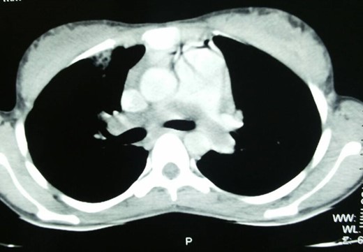
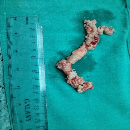
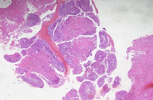
A 25-year-old lady was admitted in the pulmonology ward with the diagnosis of pneumonia. The patient had presented with a history of progressive shortness of breath, chest pain and persistent cough with occasional haemoptysis. She had a history of being treated with antibiotics and anti-TB drugs in outpatient basis. She had had spontaneous abortion 8 months back. Since her symptoms were persistent and her general condition was deteriorating, she was admitted to the ward. On examination, she had crepitations in bilateral chest and an oxygen saturation of only 80%. Chest X-ray showed bilateral infiltrations. CECT chest was done, which revealed pulmonary embolus occluding the main pulmonary artery, and right and left pulmonary arteries (Fig. 1). Venous Doppler did not reveal thrombosis of the lower limbs or IVC. Pulmonary thromboembolectomy was done and embolus (Fig. 2) was sent for histopathological examination. The examination revealed it to be metastatic choriocarcinoma (Fig. 3). Serum beta-human chorionic gonadotrophin (HCG) level was found to be significantly high. CECT pelvis and head were negative for pelvic and CNS metastases. She was managed further with chemotherapy (EMACO regimen) with excellent response to the treatment.


DISCUSSION
Choriocarcinoma presenting as pulmonary embolism is rare. Literature review identifies very few of such cases. Choriocarcinoma can metastasize to lungs, vagina, brain and liver. Women in the reproductive age group with lung metastasis present with dyspnoea, chest pain, cough and haemoptysis. As the diagnosis can always be misleading and patient might be treated in the line of pneumonia or tuberculosis, it is very essential to have a high index of suspicion. Serum beta-HCG levels are always helpful in confirming the diagnosis. Chest X-rays can show non-specific findings. CT and MRI can always provide evidence of pulmonary embolism. Surgery is indicated in patients with haemodynamic instability and those with massive tumour burden occluding main pulmonary and branch pulmonary arteries [5]. Microembolization is not amenable to surgery.
Choriocarcinomas respond very well to the chemotherapeutic agents [6]. So, chemotherapy should be initiated as soon as the diagnosis is strongly suspected or confirmed. Pulmonary embolism due to choriocarcinoma should always be suspected in a reproductive age woman presenting with intractable shortness of breath.
CONFLICT OF INTEREST STATEMENT
None declared.


