-
PDF
- Split View
-
Views
-
Cite
Cite
Erkam Komurcu, Umut Hatay Golge, Burak Kaymaz, Nilsen Erdogan, Popliteal schwannoma mimicking baker cyst: an unusual case, Journal of Surgical Case Reports, Volume 2013, Issue 8, August 2013, rjt066, https://doi.org/10.1093/jscr/rjt066
Close - Share Icon Share
Abstract
Schwannoma, also known as neurilemoma, is the most common tumour of peripheral nerves. Although it is the most common tumour of peripheral nerves, it is seldom seen in adult population. We present a very rare case of schwannoma in an unusual localization. The presented case concerns a 58-year-old patient with a slowly growing popliteal mass and neuralgia for 6 months. A mass originating from a nerve or compressing a nerve was thought in the differential diagnosis. Ultrasonographic and magnetic resonance imaging revealed a heterogenous, well-defined solid mass that seems to originate from tibial nerve. Surgical excision and histopathological examination confirmed the diagnosis of schwannoma. Diagnosis of the neurilemmoma originating from lower extremity peripheral nerves may be delayed because the mass can be misdiagnosed as baker cyst or the symptoms of the patient can be thought as a result of lumber disc herniation.
INTRODUCTION
Schwannoma, also known as neurilemoma, is the most common benign tumour of peripheral nerves originating from the schwann cells of the nerve sheath [1, 2]. Schwannomas are usually solitary, encapsulated and homogenous masses and present with slowly growing masses, sometimes associated with pain and paresthesia [1–3]. Schwannomas are usually isolated masses <2.5 cm in diameter, but the size of these tumours may differ according to localization [1]. These tumours may be located anywhere in the longitudinal axis of the extremity originating from nerve sheath of a peripheral nerve [1]. Because of their similar consistency, they may be misdiagnosed as ganglion [3]. Preoperative evaluation is based on ultrasonography and magnetic resonance imaging (MRI), but final diagnosis requires histopathology. The characteristic histological features include the presence of alternating Antoni A and Antoni B areas. Antoni A area is composed of spindle-shaped Schwann cells arranged in interlacing fascicles. There may be nuclear palisading. In-between two compact rows of well-aligned nuclei, the cell processes form eosinophilic Verocay bodies. Antoni B area consists of loose meshwork of gelatinous and microcystic tissue [4]. The mass usually appears at trunk, head and neck, or at upper extremity [5]. Localization in the lower extremity originating from tibial nerve sheath is very uncommon and rarely reported in the literature. Diagnosis of tibial nerve neurilemmoma is usually delayed because only 48% of these masses can be detected [6] and can also be misdiagnosed as Baker cyst. In this article, we present a case of popliteal schwannoma that can be misdiagnosed as Baker cyst.
CASE REPORT
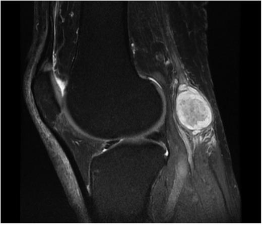
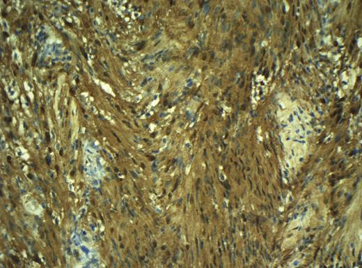
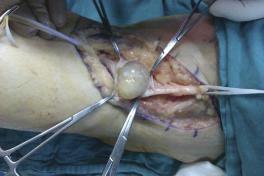
Fifty-eight-year-old man presented to the outpatient clinic of orthopaedics department with the complaints of slowly growing mass on his right popliteal region and mild pain and intermittent paresthesia of right leg and right foot. The patient emphasized that the mass existed for 2 years but has grown in the last 6 months. On physical examination, 4 × 2 cm mobile, solid mass was palpable at the posterior of the knee. There was no erythema, warmth or ulceration and Tinel's sign was positive. Sensory and motor examinations of lower extremity were normal. Ultrasound imaging showed that the mass was solid and separate from the adjacent muscles and tendons. MRI showed well-defined solid, heterogenous, dense mass originating from the tibial nerve (Fig. 1). Surgical exploration and excisional biopsy was performed. At prone position with longitudinal incision, popliteal region was explorated and tumoural mass originating from tibial nerve was dissected. Sheath of the nerve was incised longitudinally to minimize damage to the nerve fascicles and the mass was resected in en-bloc form by sharp dissection using the microscope with no complication. The patient was discharged at the post-op third day. At the third week of the operation, the patient was free of all complaints. Histopathological examination of the mass revealed hypocellular Antoni B and spindle-shaped Schwann cells containing Antoni A areas with nuclear palisading. Also immunohistochemical staining (S100+) confirmed the diagnosis of schwannoma.

DISCUSSION
Schwannoma is the most common benign neoplasm of the peripheral nerves and may originate from any of peripheral nerves. It is usually solitary, painless, encapsulated and well-defined, slowly growing mass [1–3]. In our case, tumoural mass was solitary and well-defined but painful. Tinel's sign can be important in differential diagnosis. Maraziotis et al. reported two cases of neurilemoma localized in the popliteal fossa. Both patients experienced non-specific symptoms, such as painful numbness and burning dysaesthesia, involving the lower extremity and Tinel’s sign was positive over the popliteal fossa in one of these cases [7]. In our case, Tinel's sign was positive. In the literature, tarsal tunnel syndrome secondary to tibial nerve schwannoma was reported and MR imaging was advised. Sometimes it is impossible to differentiate the schwannoma from neurofibroma or malignant peripheral nerve sheath tumours and biopsy can be a necessity to confirm the diagnosis. [3]. In our case, we also preferred to excise the tumour and confirmed the diagnosis by histopathological examination. Histopathological examination of the schwannoma reveals hypocellular Antoni B and spindle-shaped Schwann cells containing Antoni A areas with nuclear palisading. Also immunohistochemical staining (S100+) confirms the tumoural cells originating from schwann cells and the diagnosis of schwannoma [8]. In our case, necrosis, increased mytotic activity or atypical cells were not observed in histopathological examination and also positivity of S100 staining confirmed the diagnosis of schwannoma (Fig. 2 and 3). Surgical excision should be performed carefully not to damage the nerve fascicules [1]. After surgical excision, sometimes paresthesia may be seen but usually it resolves without any neurological problem and also recurrence of the tumour is very uncommon [1–3]. In this case, we did not meet any neurological complication. At the last control the patient was free of symptoms and there was no sign of residue or recurrence.


In conclusion, baker cysts are very common pathologies of popliteal region, but other benign and malignant tumours should also be kept in mind. We suggest MR investigation for differential diagnosis and excisional biopsy for confirmation of diagnosis and we also suggest excision of the schwannoma with careful dissection by using a microscope to minimize the complications and not to damage the nerve fascicules.


