-
PDF
- Split View
-
Views
-
Cite
Cite
Y Oktay, A Fikret, Leiomyosarcoma of the breast, Journal of Surgical Case Reports, Volume 2011, Issue 7, July 2011, Page 1, https://doi.org/10.1093/jscr/2011.7.1
Close - Share Icon Share
Abstract
Leiomyosarcomas of the breast are rare tumors. Less than 16 such cases have been reported in the literature so far. We present a case of a 44 year female patient who was found to have primary leiomyosarcoma of the breast.
INTRODUCTION
Stromal sarcomas of the breast constitute about 1% of all malignant tumours of the breast (1). Primary leiomyosarcoma of the breast comprises a rare type of stromal sarcoma of the breast. In the English language literature, we only found 16 similar cases described so far (2). We present a case of a 44 year old female patient, whose breast tumor had been present for several month prior to her admission.
HISTORY
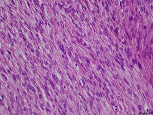
A 44 year old female patient presented to the surgical team with a firm lump in her left breast. The lesion had been present for three months when ultrasonography was performed, which found a lobulated tumor 3.5 cm in diameter in the lower in quadrant of her left breast. It was a hypoechoic and heterogeneous mass with an irregular shape, rough border and an irregular boundary echo. Fine needle aspiration biopsy was performed and was inconclusive. After discussion the multidisciplinary team decided that the patient should undergo excisional biopsy. A lumpectomy of the left breast was carried out. The subsequent histological diagnosis was stromal sarcoma with leiomyosarcomatous pattern. Microscopically, the tumor resected with mastectomy was a hypercellular nodule, and was composed of pleomorphic and hyperchromatic spindle-shaped cells arranged in an interdigitating fascicle (The tumors were circumscribed microscopically and were mild hypercellular nodules. Their nuclei were spindle shaped and showed mild to moderate atypia. Few mitoses were seen, and the surgical margins were free of tumor cells (Fig 1).

Microphotograph of leiomyosarcoma. Hematoxylin and eosin stain demonstrates a highly cellular, pleomorphic, and spindle shaped tumor with few mitotic figures (400×).
The patient underwent radiotherapy as adjuvant therapy. Twelve months later, PET-CT and whole body bone scintigraphy was clear of any recurrence.
DISCUSSION
Primary sarcoma of the breast is an unusual condition that accounts for less than 1% of all breast malignancies and less than 5% of all soft tissue sarcoma. Breast leiomyosarcomas are rare with only 16 case reports published to date (3).
Waterwarth reported the first convincing case of leiomyosarcoma of the breast in 1968 (4). Although this tumor was reported as fibrosarcoma, the tumor revealed typical light and electron microscopic features of the classic leiomyosarcoma. The mean age of the patients was 55.5 years (range, 24–86 years). The tumors tend to present as large masses, the mean size of the tumors was 4.7 cm (range, 1–9 cm). Most patients have been female but at least two of the reported cases occurred in the male breast. Nearly half of the tumors are in or near the nipple. In most cases the tumors are most likely to originate from the blood vessels or the smooth muscle of the nipple-areola complex. Most leiomyosarcomas of the breast are wellcircumscribed tumors. Microscopically, the tumors are composed of pleomorphic and hyperchromatic spindle-shaped cells arranged in an interdigitating fascicle. The cytological features reported were hyperchromasia in the nuclei, pleomorphism and mitoses. In previous literature, the mitoses of the tumors ranged from two to 21 per 10 HPF with an average of 10 mitoses (5).
Cianno et al. suggested that the presence of three mitotic figures per 10 HPF is sufficient to designate sarcomas. Nielsen has proposed that all recurring tumors that have two mitotic figures per 10 HPF should be considered leiomyosarcoma. 5 Primary leiomyosarcoma of the breast is also a very rare tumour and less than 20 well-documented cases have been reported in literature (6).
It presents as a firm lobulated mass and resembles clinically a malignant phyllodes tumour. It is difficult to establish a diagnosis of this rare tumour by fine needle aspiration cytology. Conventionally, the diagnosis of leiomyosarcoma of the breast is made on postoperative specimens by using immunohistochemical stains. The malignant spindle cells of the tumour are immunoreactive for SMA, vimentin and desmin and negative for epithelial markers and growth factor receptors (7,8).
Leiomyosarcomas of the breast are rare neoplasms that either arise from the smooth muscle cells lining blood vessels or from stromal mesenchymal cells (9). Breast sarcomas are treated by the same principles used in other sarcomas. Thus, wide local excision/lumpectomy or mastectomy with or without radiotherapy are used. Lym-phatic spread and nodal metastasis are not features associated with these neoplasms. Axillary nodal metastasis occurs in less than 10% of breast sarcomas, making sentinel lymph node biopsy or axillary lymph node dissection unnecessary (10).
Leiomyosarcoma is a rare malignant tumor of the breast. Surgical treatment is the mainstay of therapy. Axillary dissection, chemotherapy, and radiotherapy have not been shown to improve the rates of disease-free or overall survival. Because of the risk of late recurrence, long-term follow-up is necessary.


