


Abstract
Giant inguinal herniae pose a surgical challenge, though not uncommon in the developing world they are a rare presentation in the UK. We present a patient with cardiac disease who presented with a giant inguino-scrotal hernia complicated by a bleeding scrotal ulcer. We describe his medical management and the surgical repair of the hernia and refashioning of his scrotum.
INTRODUCTION
A giant inguino-scrotal hernia is defined as a hernia that extends below the midpoint of the inner thigh in the standing position (1). Most patients would have had their hernia for several years. Complications include impairment of mobility (2, 3, 4), incarceration and intestinal obstruction (3). Scrotal ulceration (5) is also common; this is due to pressure necrosis or frictional rub. Other presenting features include as scrotal elephantiasis and gangrene (6). This type of hernia poses a major surgical challenge and the operative approach requires careful planning.
CASE REPORT
A 52 year old history teacher presented as an emergency with a recurrent bleeding scrotal ulcer. Three months previously he had undergone a double cardiac valvular replacement for severe aortic and mitral valvular disease following an attack of rheumatic carditis as a child. He was placed on Warfarin post-operatively. The patient had had a right inguinal hernia for 20 years but was deemed unfit for surgery because of his diseased cardiac valves; he had to give up work as his hernia grew to a size where he could barely walk.
On examination he was hemodynamically stable. Abdominal examination revealed a giant right inguino-scrotal hernia; there was a large 15 by 15 cm ulcer on the base of the right hemiscrotum that was bleeding. The penis was engulfed in the swelling (fig 1). His haemoglobin was 8g/dl and his INR 3.9.
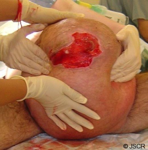
The giant inguinoscrotal hernia demonstrating the scrotal ulcer
The scrotal wound was dressed and the patient was transfused. His INR was reversed and under the cover of antibiotics he underwent a Lichenstein repair of his right inguino-scrotal hernia through a right groin incision. At operation the hernia sac contained the entire length of the small and large intestine, the appendix was stuck to the base of the scrotum and the stomach was in the pelvis. Four litres of straw coloured fluid was drained from the hernia sac. The intestine was reduced out of the scrotum, an appendicectomy was performed and the contents replaced back into the abdomen. The patient was placed in the Tredenlenburg position. The cord structures were transected in the groin. The sac was closed with 2/0 vicryl, transveraslis fascia was plicated and a prolypropylene mesh was used to repair the posterior wall of the inguinal canal. A vertical right hemiscrotal incision was made the right testis was delivered, the redundant skin and ulcer was excised. The dartos muscle was closed with interrupted 0 vicryl and the skin with haemostatic 2/0 vicryl rapide mattress sutures. A mini-circumcision had to be performed to enable urethral catherisation.
Post-operatively the patient was transferred to the intensive care unit, was extubated two hours after surgery and did not suffer any respiratory problems. He was recommenced on heparin perioperatively and Warfarin 48 hours post-operatively. He was discharged home 6 days later with an INR of 3.2. He was re-admitted with bleeding from his mini-circumcision wound on the 12th post-operative day, his INR was 5.9, this was corrected and he was discharged 5 days later on warfarin.
He was reviewed in the clinic 12 weeks later, he was satisfied with the final outcome (fig 2) and was about to return to work.
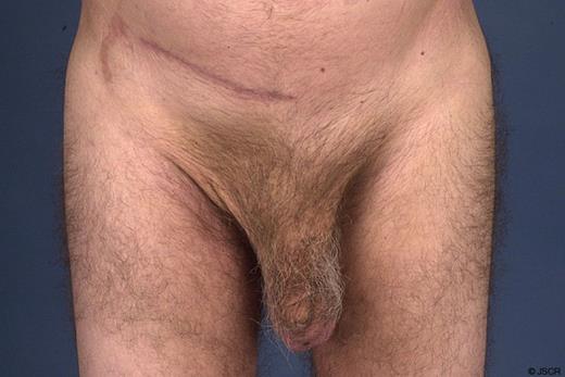
Image taken 3 months postoperatively
DISCUSSION
This case illustrates the problems patients may encounter with a giant inguinoscrotal hernia. Our patient had been suffering for many years and had given up his job due to resulting disability. He presented with a bleeding scrotal wound that was compounded by the fact that he was on Warfarin.
The surgical management of a patient with a giant inguino-scrotal hernia should be carefully planned. The key factors to consider are the operative approach, whether or not to use a mesh for the repair, how to deal with the sudden increase in intra abdominal pressure due to loss of domain, obtaining informed consent and dealing with the redundant scrotal skin or scrotal haematoma. We had the added challenge of ensuring that our patient did not develop infective endocarditis as well as tightly controlling his clotting to prevent post-operative bleeding and thrombosis.
The operative approach could be through an inguinal incision, laparotomy or as a combined approach (3). We chose an inguinal approach in our patient for recovery from a laparotomy would have taken longer and he had to be re-anticoagulated immediately after surgery. If the patient is unstable or has a strangulated hernia the operation may have to be performed in stages (5, 6). If the operation is going to be performed in one stage careful consideration should be paid to the type of implant used to repair the hernia defect, it is inadvisable to use a mesh if a bowel resection is performed. There are tissue grafts that could be used instead, these are expensive and may not be readily available for emergency use.
Giant inguinal herniae tend to be commoner in males hence the patient should be consented for an orchidectomy. A testicular prosthesis could be inserted as a second procedure.
Most patients with giant inguino-scrotal herniae tend to have most of their abdominal contents sitting in the scrotum (3, 7), as result, the abdominal cavity would have shrunk; sudden replacement of these contents back into the abdominal cavity could lead to respiratory embarrassment. Various modalities have been tried to deal with this problem. In a developed country where intensive care facilities are readily available the hernia contents could be reduced into the abdominal cavity and the patient could be ventilated until they are able to breath unaided, this was the approach taken in our patient. Such patients should be carefully monitored for signs of intra abdominal compartment syndrome. In developing countries where medical facilities are limited creating a pneumoperitoneum over a number of days to re expand the abdominal cavity is the desired option (2, 8, 9) however, there is a risk of infection. Creating a pneumoperitoneum does not always lead to re-expansion of the abdominal cavity especially if the air leaks into the hernia sac. Other surgeons have made relaxing incisions in the flanks to increase the abdominal girth (4, 10) or placed an interposition mesh in the abdominal wound (4, 6, 10). Mehendal et al performed a bowel resection in order to reduce the amount of viscera reduced back into the abdomen; this approach is difficult to justify in a patient with viable bowel for creating an anastomosis adds to the post-operative risks.
Once the hernia has been dealt with the redundant scrotal skin should be addressed as a primary or delayed procedure depending on the clinical status of the patient (1,4). In our patient the indication for surgery was haemorrhage from a scrotal ulcer so the redundant skin and the ulcer were excised. One should exercise caution in excising the scrotal skin for the skin is usually oedematous and is likely to shrink post operatively (3, 4). If the redundant scrotal skin is not excised there is a higher risk of haematoma formation, which could take several weeks to resolve. Our patient had to be anticoagulated 8 hours after surgery, haemostasis was ensured intra-operatively and he did not develop a haematoma.
