


Abstract
Congenital bladder diverticula in children are uncommon and rarely present with bladder outlet obstruction. We present a case highlighting an interesting association between a congenital bladder diverticulum and a benign inflammatory bladder wall lesion mimicking a rhabdomyosarcoma. Open surgery was required as different imaging modalities and cystoscopy were insufficient to exclude a malignant process.
INTRODUCTION
Rhabdomyosarcoma of the bladder in children is a rare but highly aggressive malignancy. It is often an occult condition that presents with bladder outlet obstruction or haematuria. The authors present a case of congenital bladder diverticulum and a benign inflammatory bladder wall lesion mimicking bladder rhabdomyosarcoma. Open surgery was required to rule out malignant pathology despite different imaging modalities.
CASE REPORT
A previously well 2-year old boy presented with acute retention of urine requiring catheterisation. Renal tract ultrasonography revealed a large right-sided bladder diverticulum and a 2.2 x 1.3 x 3.6 cm lobulated thickening on the posterior bladder wall, which was associated with increased vascularity on Doppler imaging (Figure 1).
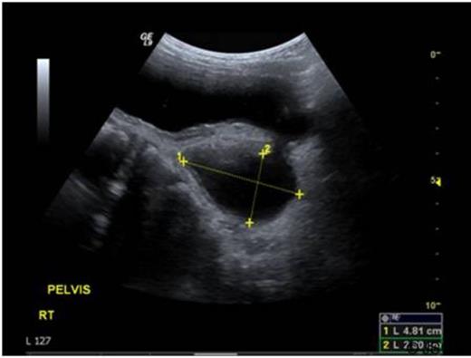
Ultrasound images demonstrating a large bladder diverticulum
The diverticulum was clearly seen on micturating cystourethrogram (MCUG). Subsequent magnetic resonance imaging (MRI) scan confirmed the presence of a large bladder diverticulum and also demonstrated an enhancing, irregular, nodular posterior bladder wall thickening which was confined to the bladder. Cystoscopy revealed a papillary lesion on the posterior bladder wall adjacent to the diverticulum opening (figure 2).
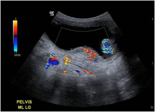
Ultrasound images demonstrating a posterior bladder wall lesion with increased vascularity
The cystoscopic appearances resembled a rhabdomyosarcoma. Endoscopic biopsies demonstrated no evidence of malignant change. Diverticulectomy and ureteric re-implantation were performed to resolve bladder outlet obstruction and an open bladder biopsy of the lesion was conducted at the same time. Histological appearances were consistent with polypoidal cystitis. Immunohistochemistry was negative on conventional immunostains for rhabdomyosarcoma (i.e. desmin and cytokeratin). The patient made an uneventful recovery and continues to do well with no further episodes of urine retention at outpatient review.
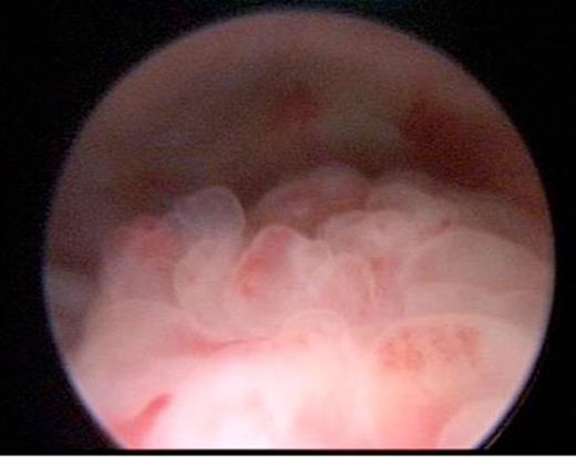
Papillary lesion seen on posterior bladder wall at cystoscopy
DISCUSSION
Congenital bladder diverticula in children are uncommon and rarely cause bladder outlet obstruction (1–2). Rhabdomyosarcomas are rare entities that frequently present with urinary retention. Benign inflammatory lesions can mimic rhabdomyosarcomas both radiologically and macroscopically as in the present case (3). Histological examination of bladder wall lesions is therefore recommended to establish diagnosis. Corbett et al reported an identical case of congenital bladder diverticulum co-existing with a benign lesion resembling a tumour (4). Based on the present case and the case reported by Corbett et al, the authors postulate that benign inflammatory lesions mimicking tumours occur with congenital bladder diverticula but due to their transient nature are not frequently reported. In cases where dual pathology of bladder wall lesions and diverticula are observed, clinicians can be reassured that this is probably a benign process.
