


Abstract
Amputation is frequently required to treat wet gangrene secondary to peripheral vascular disease. Although different types of amputations have been reported, limited digital and ray amputation are the commonest amputations performed. The level of amputation will be determined by the distribution of lower limb disease but for every patient a balance between limb and functional preservation at lower levels versus better wound healing at higher levels should be sought.
In this article we describe the novel osteo-myocutaneous flap, which we have used in our patient. We believe that this flap results in improved wound healing and although it results in loss of two digits, it does not impair function.
INTRODUCTION
Different types of amputation are performed for wet gangrene secondary to peripheral vascular disease. We describe the novel osteo-myocutaneous flap in a 64 year old man.
CASE REPORT
Our patient, a 64 year old gentleman presented via the Emergency Department with wet gangrene of the right fourth toe, associated with a severe cellulitis affecting the distal third of the right lower limb. Lower limb pulses were palpable with the exception of the right tibialis posterior artery. The patient had been treated in the community for ulcers of mixed aetiology over the right lateral malleolus and heel. Co-morbidities included MRSA colonisation, NIDDM, hypertension and peripheral vascular disease, the latter of which had resulted in left below knee amputation three years prior.
Conservative management was instituted, including intravenous rehydration, antibiotics (Vancomycin and Metronidazole), with MRI angiogram assessment demonstrating mild disease of the right external iliac artery and severely diseased right-sided arterial runoff below the trifurcation. Following initial improvement secondary to intravenous antibiotics, no further progress was noted and resolution of cellulitis suggested involvement of the third and fifth digits. Consequently, the patient consented to amputation of his fourth toe +/- the third and fifth digits. The patient also consented to below knee amputation if this was deemed necessary intra-operatively.
The procedure was performed under regional anaesthesia, with a racket incision centred over the fourth metatarsophalangeal joint necessary to excise the fourth digit. The incision began 1 cm proximal to the metatarsophalangeal joint and passed distally to the base of the proximal phalanx which was divided to pass around the toe and across the plantar surface at the level of the flexor crease. The amputated segment was removed and sent to the laboratory for microbiological microscopy, culture and sensitivity analyses. The skin of the third and fifth toes adjacent to the fourth toe was debrided as necessary, although no further amputation was required. We opted to close this wound by constructing a tissue flap rather than leaving the wound to granulate by secondary intention. In this instance we utilised the fifth digit as a novel osteo-myocuteanous flap, rather than using conventional techniques. As can be seen from the x-ray studies performed postoperatively, we modified standard myocutaneous flap construction techniques by incising the medial border of the fifth digit to the periosteum, which was then mobilised to the superior / inferior surface of the fifth phalanxes, with the bone remaining in-situ (Figure 1).

Postoperative x-ray of the right foot demonstrating the amputated fourth toe. The Xray also demonstrates the phalanxes of the fifth toe being used to construct the flap used to close the defect.
The flap was sutured to the surrounding tissue using non absorbable 3,0 Prolene sutures. The patient made an uneventful recovery and following resolution of the cellulitis and after social services input he was discharged nine days postoperatively. Figures two and three demonstrate the appearance of the flap on postoperative day seven.
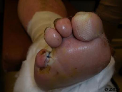
Close-up appearance of the flap seven days postoperatively.
DISCUSSION
Amputation is frequently required to treat wet gangrene secondary to peripheral vascular disease, with numerous procedures reported including: limited digital, ray, transmetatarsal, and hindfoot amputations. Ultimately, the level at which amputation must occur will be determined by the distribution of lower limb disease. Nevertheless, good preoperative assessment is mandatory for each individual patient presenting with a gangrenous foot, to balance limb preservation following limited distal amputation against the superior wound healing associated with above/below knee amputation.
While the psychological and physical benefits associated with limb preservation are well established, conventional procedures are not without complication. Procedures performed for diabetic patients are generally either limited digital or ray amputations. The wound generated by amputation of a single toe is usually either closed primarily or left to heal by secondary intention, although both strategies are frequently impaired by diabetic vascular disease. This complication may be more troublesome following ray amputation since the skin defect and tissue loss is more extensive. In contrast, this report suggests that the use of an osteo-myocuteanous flap may result in effective wound healing following limited digital amputation in the setting of diabetic microvascular disease, presumably due to increased vascularity at the wound site. While the use of an osteo-myocutaneous flap will result in loss of two digits, this is often necessary to adequately remove gangrenous tissue and does not cause any disturbance in function. The results in this patient have been good and no complications were observed within the first 30-day period postoperatively. Consequently, we believe this technique may be of great benefit to those patients for whom single toe amputation may not have been considered as healing might have been problematic. However, we do acknowledge that toes amputated using this technique may still be susceptible to the complications that may occur in toe amputations using the conventional technique. These include deviation of the remaining toes to fill the gap left by the amputation thereby altering the biomechanics.(1) This risk is increased in amputations involving the second toe and may result in a hallux valgus deformity. This may ensue in inadvertent pressure areas that may necessitate further amputations. Further prospective randomised studies are thus required to compare this procedure with conventional techniques, to confirm the efficacy of osteo-myocuteanous flap construction and other potential postoperative complications.
